HYBRID EVENT: You can participate in person at Tokyo, Japan from your home or work.
Echocardiography, Cardiac CT and MRI
Echocardiography, Cardiac CT and MRI
Echocardiography: An echocardiography is a test that uses high frequency sound waves to produce live images of heart to see structure of heart and to evaluate heart functions. The image is called an echocardiogram. It has no side effects. Echocardiography helps doctor to find out:
- The size and shape of heart, thickness and movement of heart’s wall.
- How heart moves
- The heart’s pumping strength.
- If the heart values are working correctly.
- If blood is leaking backwards through heart values.
- If the heart values are too narrow
- If there is a tumor or infectious growth around heart values
Cardiac CT: Cardiac CT is also known as computed tomography of heart and is performed to know about cardiac anatomy to diagnose coronary artery disease, to check patency of coronary artery bypass grafts or to assess volume try and cardiac function.
MRI: MRI uses radio waves and a strong magnetic field to create images of organs and tissues with the body. MRI scan varies from CT scans and X-rays as it does not involve use of potentially harmful ionizing radiations.
Committee Members

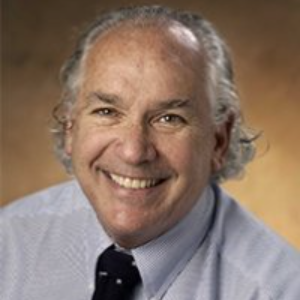
Arthur J Siegel
Massachusetts General Hospital, United States
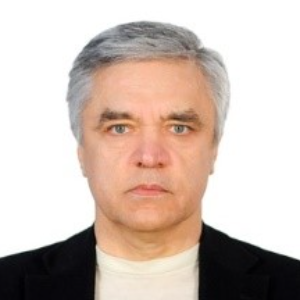
Sergey Suchkov
N. D. Zelinskii Institute for Organic Chemistry of the Russian Academy of Sciences, Russian Federation
Narendra Kumar
HeartbeatsZ Academy, United Kingdom Cardio 2026 Speakers

Arthur J Siegel
Massachusetts General Hospital, United States
Yong Xiao Wang
Albany Medical Center, United States
Narendra Kumar
HeartbeatsZ Academy, United Kingdom


Title : New recommendations for the prevention of sudden cardiac death in athletes and recreational sports
Sekib Sokolovic, ASA Hospital Sarajevo, Bosnia and Herzegowina
Title : Coronary revascularization in patients with diabetes: Prospects for stenting in patients with type 1 diabetes and coronary artery disease
Mekhman N Mamedov, National Research Center for Therapy and Preventive Medicine, Russian Federation
Title : An adult case of polysplenia syndrome associated with sinus node dysfunction
Apoorva Tripathi, Oxford University Hospitals, United Kingdom
Title : Personalized and precision medicine (PPM) as a unique healthcare model through biodesign-driven translational applications and cardiology-related healthcare marketing to secure the human healthcare and biosafety
Sergey Suchkov, N. D. Zelinskii Institute for Organic Chemistry of the Russian Academy of Sciences, Russian Federation
Title : A unique cell-driven phenomenon in the heart and the promising future of the innovative translational tools to manage cardiac self-renewal and regeneration
Sergey Suchkov, N. D. Zelinskii Institute for Organic Chemistry of the Russian Academy of Sciences, Russian Federation
Title : Young hearts at risk: Hidden cardiovascular damage and the role of social determinants of health among youth with type 1 diabetes in Kenya
Phoebe Wamalwa, Ministry of Health, Kenya